Histology Of Smooth Muscle Diagram - Skeletal muscle is one of the three types of muscle tissue, alongside cardiac and smooth muscle.
byAdmin-
0
Histology Of Smooth Muscle Diagram - Skeletal muscle is one of the three types of muscle tissue, alongside cardiac and smooth muscle.. Muscle, one of the four basic tissues of the body, is specialized for contraction. Development of skeletal muscle, cardiac muscle and smooth muscle can be found in other notes. 102 skeletal muscle anatomy and physiology. The juxtaglomerular cells, derived from smooth muscle cells, of the afferent arteriole secrete renin when blood pressure in the arteriole falls. It attaches to bones and the orbits through tendons.
2 energy sources for muscles. Labelled diagram of a muscle cell places to visit muscle. Development of skeletal muscle from mesoderm occurs by mononucleated myoblasts. Smooth muscle has inherent contractility, and the autonomic nervous system, hormones and local metabolites can influence its contraction. Four basic types of tissues are found in animals.
Histology 4000 Muscle Lecture Notes 6 from www.auburn.edu Smooth muscle is in the lower panel. This diagram shows a skeletal muscle cell it labels the dark and. A diagram of a muscle sarcomere is shown below. It also occurs in the spleen (capsule and trabeculae), eye (iris and ciliary body), skin (arrector pili muscles of hairs), endocardium, scrotum, penis. This article will discuss the structure of skeletal muscle tissue, its mode of contraction and relevant clinical conditions. Myosin 2 is composed of. Steps of sliding filament theory of contraction. Excitable tissue responds to stimuli through electrical signals.
In this short guide, you will get a basic concept of skeletal muscle histology from the real slide and labeled diagram.
The glandular tissue is made up of branching tubuloalveolar glands and the stroma consists of smooth muscle, collagen, elastic tissue and fibrous tissue. This article will discuss the structure of skeletal muscle tissue, its mode of contraction and relevant clinical conditions. It appears smooth since the protein filaments are not arranged regularly in order. Smooth muscle, muscle that shows no cross stripes under microscopic magnification. Introduction to histology (part 1) tissues are composed of similar types of cells that work in a coordinated fashion to perform a common task, and the study of the tissue level of biological organization is histology. Ninja nerds, join us in this video where we discuss the histology, anatomy, and function of smooth muscle within the human body.***please support us***patreo. Phosphagen energy system (with atp and creatine. Since it is not under conscious. At the lower one third of ureter histology of animal, you might find another smooth muscle layer. It is classified as a striated muscle tissue, which functions to contract and permit movements under voluntary control. Labelled diagram of a muscle cell places to visit muscle. In this short guide, you will get a basic concept of skeletal muscle histology from the real slide and labeled diagram. The cells are spindle shaped, and the nucleus is central.
Muscle histology of muscle and bone type skeletal cardiac smooth nucleus multinucleated; • smooth muscles respond to stretch only briefly, and then adapts to its new length. The skeletal muscle fibers are elongated, cylindrical and multinucleated cells whose length may vary in different animals. A band remains the same size, i and h bands reduce in size. At the lower one third of ureter histology of animal, you might find another smooth muscle layer.
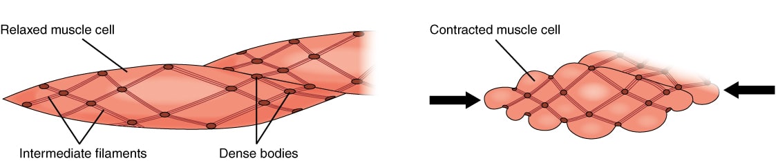
Smooth Muscle Tissue Structure Contraction Teachmephysiology from teachmephysiology.com This diagram shows a few of the cells that can be seen in the stained section below. Neural impulse on sarcolemma causes ach release and depolarization. Steps of sliding filament theory of contraction. The glandular tissue is made up of branching tubuloalveolar glands and the stroma consists of smooth muscle, collagen, elastic tissue and fibrous tissue. Development of skeletal muscle from mesoderm occurs by mononucleated myoblasts. Phosphagen energy system (with atp and creatine. It is found in the walls of ducts and blood and lymphatic vessels, as well as in the walls of the digestive, respiratory and urogenital tracts. The cells stick together and are connected by specialised cell junctions, called gap junctions.
This diagram shows a few of the cells that can be seen in the stained section below.
Cardiac muscle is in the center; Microscopy and the study of tissues. Smooth muscle has inherent contractility, and the autonomic nervous system, hormones and local metabolites can influence its contraction. The cells are spindle shaped, and the nucleus is central. Development of skeletal muscle from mesoderm occurs by mononucleated myoblasts. It is composed of 30% glandular tissue (white) and 70% stromal tissue (pink). Each muscle fiber has oval nucleus in the center. Skeletal muscle is an excitable, contractile tissue responsible for maintaining posture and moving the orbits, together with the appendicular and axial skeletons. Ca2+ pumps in membrane of sarcoplasmatic reticulum actively drive ca2+ ions back into terminal cistaern where it's bound to calsequestrin. At the lower one third of ureter histology of animal, you might find another smooth muscle layer. In response to elevated sodium, the macula densa cells trigger contraction of the afferent arteriole, reducing flow of blood to the glomerulus and the glomerular filtration rate. Spindle shaped, non striated, involuntary muscle fibers. But sometime these two layers of smooth muscle bundles are difficult to distinguish in ureter of few animals.
This article will discuss the structure of skeletal muscle tissue, its mode of contraction and relevant clinical conditions. In this short guide, you will get a basic concept of skeletal muscle histology from the real slide and labeled diagram. Development of skeletal muscle from mesoderm occurs by mononucleated myoblasts. Neural impulse on sarcolemma causes ach release and depolarization. 2 energy sources for muscles.
Equine Duodenum Histology Medical Laboratory Science Smooth Muscle Histology Slides from i.pinimg.com Skeletal muscle is an excitable, contractile tissue responsible for maintaining posture and moving the orbits, together with the appendicular and axial skeletons. Cardiac muscle is in the center; It is classified as a striated muscle tissue, which functions to contract and permit movements under voluntary control. Steps of sliding filament theory of contraction. At the lower one third of ureter histology of animal, you might find another smooth muscle layer. Phosphagen energy system (with atp and creatine. It is found in the walls of ducts and blood and lymphatic vessels, as well as in the walls of the digestive, respiratory and urogenital tracts. In this short guide, you will get a basic concept of skeletal muscle histology from the real slide and labeled diagram.
This diagram shows a few of the cells that can be seen in the stained section below.
Development of skeletal muscle from mesoderm occurs by mononucleated myoblasts. Smooth muscle is widely distributed in the body. Excitable tissue responds to stimuli through electrical signals. This diagram shows a few of the cells that can be seen in the stained section below. Four basic types of tissues are found in animals. Skeletal muscle is an excitable, contractile tissue responsible for maintaining posture and moving the orbits, together with the appendicular and axial skeletons. Myosin 2 is composed of. First step of muscle relaxation. 2 heavy chains and 4 light chains. Smooth muscle is specialized for slow and sustained contractions of low force. A band remains the same size, i and h bands reduce in size. Muscle, one of the four basic tissues of the body, is specialized for contraction. The cells stick together and are connected by specialised cell junctions, called gap junctions.
Skeletal muscle is an excitable, contractile tissue responsible for maintaining posture and moving the orbits, together with the appendicular and axial skeletons smooth muscle diagram. Excitable tissue responds to stimuli through electrical signals.